40 diagram of the human eye without labels
Diagram of the Eye - Lions Eye Institute To understand the eye and its functions, it's important to understand how the eye works, see below diagrams for both the external eye and the internal eye. The External Eye Instructions Click the parts of the eye to see a description for each. Hover the diagram to zoom. The Internal Eye Instructions Structure and Functions of Human Eye with labelled Diagram Human Eye Diagram: Contrary to popular belief, the eyes are not perfectly spherical; instead, it is made up of two separate segments fused together. Explore: Facts About The Eye To understand more in detail about our eye and how our eye functions, we need to look into the structure of the human eye.
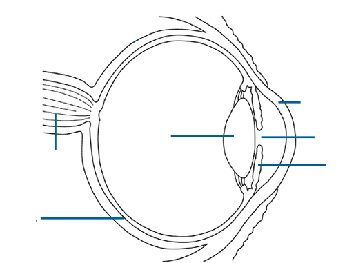
39 diagram of the human eye without labels Human eye - Wikipedia Schematic diagram of the human eye. It shows a horizontal section through the right eye. The eye is made up of three coats, or layers, enclosing various anatomical structures. The outermost layer, known as the fibrous tunic, is composed of the cornea and sclera, which provide shape to the eye and support the deeper structures.

Diagram of the human eye without labels
Human Eye Anatomy - Parts of the Eye ... - All About Vision Eye anatomy: A closer look at the parts of the eye. By Liz Segre. When surveyed about the five senses — sight, hearing, taste, smell and touch — people consistently report that their eyesight is the mode of perception they value (and fear losing) most. Despite this, many people don't have a good understanding of the anatomy of the eye, how ... The Human Eye | Boundless Physics - Lumen Learning The fundus is on the opposite of the pupil, but inside the eye and can not be seen without special instruments. The optic nerve is what conveys the signals of the eye to the brain. is a diagram of the eye. The human eye is made up of three coats: Diagram of the Human Eye: The cornea and lens of an eye act together to form a real image on the ... Eye Diagram Teaching Resources | Teachers Pay Teachers Anatomy of the Eye Diagrams for Coloring/Labeling, with Reference and Summary by Homemade For Play 7 $1.95 PDF This printable contains 13 clear and simple cross sectional diagrams of the human eye.
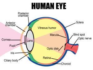
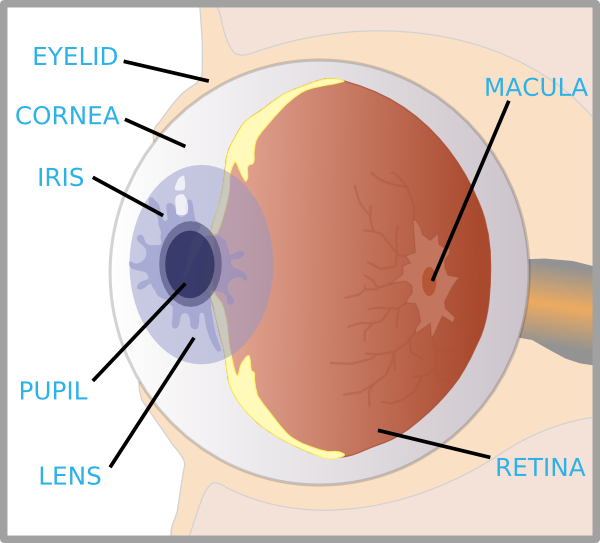
Diagram of the human eye without labels. Human eye anatomy Images, Stock Photos ... - Shutterstock Human eye anatomy royalty-free images 60,203 human eye anatomy stock photos, vectors, and illustrations are available royalty-free. See human eye anatomy stock video clips Image type Orientation Sort by Popular Biology Healthcare and Medical Icons and Graphics Recreation/Fitness human eye anatomy 3d rendering eye visual perception infographic Next File:Diagram of human eye without labels.svg - Wikimedia ... File:Diagram of human eye without labels.svg. Size of this PNG preview of this SVG file: 410 × 430 pixels. Other resolutions: 229 × 240 pixels | 458 × 480 pixels | 732 × 768 pixels | 976 × 1,024 pixels | 1,953 × 2,048 pixels. Anatomy of the eye: Quizzes and diagrams | Kenhub Take a look at the diagram of the eyeball above. Here you can see all of the main structures in this area. Spend some time reviewing the name and location of each one, then try to label the eye yourself - without peeking! - using the eye diagram (blank) below. Unlabeled diagram of the eye PDF Eye Anatomy Handout - National Eye Institute of light entering the eye. Lens: The lens is a clear part of the eye behind the iris that helps to focus light, or an image, on the retina. Macula: The macula is the small, sensitive area of the retina that gives central vision. It is located in the center of the retina. Optic nerve: The optic nerve is the largest sensory nerve of the eye.
Eye Diagram - Differentiated Worksheets and ... - Pinterest Eye Diagram - Differentiated Worksheets and EASEL Activities Description Use these simple eye diagrams to help students learn about the human eye. Three differentiated worksheets are included: 1. Write the words using a word bank 2. Cut and paste the words 3. Human eye - Wikipedia The human eye is a sensory organ, ... Schematic diagram of the human eye. It shows a horizontal section through the right eye. ... Right eye without labels ... FREE! - Label the Eye Worksheet - Teacher-Made Learning ... The first page is a labelling exercise with two diagrams of the human eye. One is a view from the outside, and the other is a more detailed cross-section. On the second page, you'll find a set of answers showing the properly labelled human eyes, designed to help you check the worksheets without having to come up with your own answer key. The Eyes (Human Anatomy): Diagram, Optic Nerve, Iris ... Articles On Eye Basics. Your eye is a slightly asymmetrical globe, about an inch in diameter. The front part (what you see in the mirror) includes: Iris: the colored part. Cornea: a clear dome ...
Human Eye - Definition, Structure, Function, Parts, Diagram A human eye is roughly 2.3 cm in diameter and is almost a spherical ball filled with some fluid. It consists of the following parts: Sclera: It is the outer covering, a protective tough white layer called the sclera (white part of the eye). Cornea: The front transparent part of the sclera is called cornea. Light enters the eye through the cornea. Success Essays - Assisting students with assignments online Get 24⁄7 customer support help when you place a homework help service order with us. We will guide you on how to place your essay help, proofreading and editing your draft – fixing the grammar, spelling, or formatting of your paper easily and cheaply. Human Body Diagram - Bodytomy Walnuts, pound for pound, resemble the human brain. The folds and crevices, too, of the brain are mimicked perfectly by nature. Rich in omega-3 fatty acids, regular consumption of this nut facilitates the functions of the brain. Carrots ⇆ Eyes Health care providers perpetually recommend to nibble on carrots to keep your eyes healthy and active. File:Schematic diagram of the human eye en.svg - Wikimedia File:Schematic diagram of the human eye en.svg. Size of this PNG preview of this SVG file: 416 × 423 pixels. Other resolutions: 236 × 240 pixels | 472 × 480 pixels | 755 × 768 pixels | 1,007 × 1,024 pixels | 2,014 × 2,048 pixels.
File:Diagram of human eye without labels.svg - Wikimedia Commons
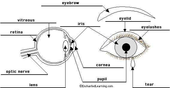
Eyes - Layers of Learning | Human eye diagram, Parts of ... Elementary Science. Description Use these simple eye diagrams to help students learn about the human eye. Three differentiated worksheets are included: 1. Write the words using a word bank 2. Cut and paste the words 3. Write the words without a word bank Labels include: eyebrow, eyelid, eyelashes, pupil, iris, and sclera.

Pin on Drawings - Drapery & Perspective sight
Draw a labeled diagram of human eye. Write the functions ... Cornea of the eye is the first sight where convergence of light rays takes place. Iris is that part of the eye which controls the amount of light entering the eye through the pupil. Pupil is a type of small hole through which light enters the eye. The eye lens is a convex lens just behind the pupilwhich converges the light rays towards the ratina. The retina is a type of screen in the eye ...

31 Human Heart To Label - Labels Design Ideas 2020
Label the Eye Diagram - Enchanted Learning Label the Eye Diagram. Human Anatomy. Read the definitions, then label the eye anatomy diagram below. Cornea - the clear, dome-shaped tissue covering the front of the eye. Iris - the colored part of the eye - it controls the amount of light that enters the eye by changing the size of the pupil. Lens - a crystalline structure located just behind ...

82 best Anatomy Physiology images on Pinterest | Nursing schools, Nursing students and Schools ...
The Eye - diagram to label | Teaching Resources File previews. pdf, 2.94 MB. Diagram of eye with key words to use in labelling it. Tes classic free licence.
Human Eye anatomy : How the Human Eye Works
Eye Anatomy: 16 Parts of the Eye & Their Functions The lens of the eye (or crystalline lens) is the transparent lentil-shaped structure inside your eye. This is the natural lens. It is located behind the iris and to the front of the vitreous humor (vitreous body). The vitreous humor is a clear, colorless, gelatinous mass that fills the gap between the lens and the retina in the eye.

Human Body Muscles Diagram Labeled : muscles to label - Google Search | Muscular system, Human ...
Eye Anatomy: Parts of the Eye and How We See - American ... Here is a tour of the eye starting from the outside, going in through the front and working to the back. Eye Anatomy: Parts of the Eye Outside the Eyeball. The eye sits in a protective bony socket called the orbit. Six extraocular muscles in the orbit are attached to the eye. These muscles move the eye up and down, side to side, and rotate the eye.
structure of eye without label - Clip Art Library
43 diagram of the human eye without labels 43 diagram of the human eye without labels May 16, 2022 Human Eye - Definition, Structure, Function, Parts, Diagram A human eye is roughly 2.3 cm in diameter and is almost a spherical ball filled with some fluid.

whatisscience: August 2010
Eye Diagram Unlabelled - Wiring Diagram Pictures Best Human eye diagram unlabelled free vector download for commercial use in ai, eps, cdr, svg vector illustration graphic art design format. human eye. Ask A Biologistcoloring page | Web address:schematron.org coloring. Human Eye. Page 2. 5. 3. 2. 4. How to draw human eye in easy steps -10th -Physics - science - CBSE syllabus - NCERT class 10
picture front of the eye without labels clipart - Clipground
Human Ear Diagram - Bodytomy Look no further, this Bodytomy article gives you a labeled human ear diagram and also explains the functions of its different components. The human body is like a big machine, and various processes take place inside it. With the help of the various organs and tissues, it carries out some of the most marvelous tasks, that are no less than a ...
Simple Labeled Human Eye Diagram - Aflam-Neeeak
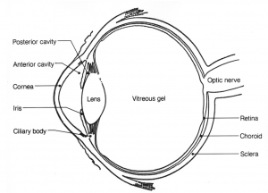
PDF Parts of the Eye - National Eye Institute Eye Diagram Handout Author: National Eye Health Education Program of the National Eye Institute, National Institutes of Health Subject: Handout illustrating parts of the eye Keywords: parts of the eye, eye diagram, vitreous gel, iris, cornea, pupil, lens, optic nerve, macula, retina Created Date: 12/16/2011 12:39:09 PM

Draw a neat and label diagram of human eye and explain functions | Meritnation.com
Label Parts of the Human Eye - University of Dayton Label Parts of the Human Eye. Select One Anterior Chamber Ciliary Body Cornea Fibrous Tunic Iris Lateral Rectus Muscle Lens Medial Rectus Muscle Optic Disk Optic Nerve Pupil Retina Vascular Tunic Vitreous Nerve.

Human eye diagram stock vector. Illustration of aqueous - 45971931
Eye Diagram Teaching Resources | Teachers Pay Teachers Anatomy of the Eye Diagrams for Coloring/Labeling, with Reference and Summary by Homemade For Play 7 $1.95 PDF This printable contains 13 clear and simple cross sectional diagrams of the human eye.
Eye With Labels Clip Art at Clker.com - vector clip art online, royalty free & public domain
The Human Eye | Boundless Physics - Lumen Learning The fundus is on the opposite of the pupil, but inside the eye and can not be seen without special instruments. The optic nerve is what conveys the signals of the eye to the brain. is a diagram of the eye. The human eye is made up of three coats: Diagram of the Human Eye: The cornea and lens of an eye act together to form a real image on the ...
NCERT Solutions for Class 8 Science Chapter 16 - Light - Arinjay Academy
Human Eye Anatomy - Parts of the Eye ... - All About Vision Eye anatomy: A closer look at the parts of the eye. By Liz Segre. When surveyed about the five senses — sight, hearing, taste, smell and touch — people consistently report that their eyesight is the mode of perception they value (and fear losing) most. Despite this, many people don't have a good understanding of the anatomy of the eye, how ...
Blank Eye Diagram - Cliparts.co
Post a Comment for "40 diagram of the human eye without labels"